
The research program is based on the three related subjects: (1) the fundamental study of light-metal interaction at nanoscale, (2) vibrational spectroscopy of individual molecules, and (3) development of new spectro-nanoscopy techniques both in visible and IR frequencies. These three topics are not only the important research projects on their own, but they also serve as the “tools” for the other two: the plasmonics forms the physical basis of the vibrational spectroscopy of individual molecules and optical nanoscopy. The vibrational spectroscopic signatures of the individual molecules serve as probes of plasmons of nanostructures. The nanoscopy provides experimental means to directly study the plasmons of individual nanostructures and also enables us to spectroscopically observe the individual molecules at nanometric resolutions.
Metals: nanoparticle plasmonics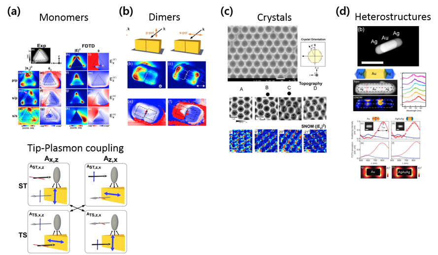
We have been studying the localized surface plasmons (LSPs) of individual Ag and Au nanoparticles and their close-coupling. To better understand the LSPs, we probe the plasmons in spatial domain (i. e., plasmon modes) as well as in frequency domain (i. e., resonance frequencies). In particular, we have shown that the scattering-type scanning near-field optical microscopy (sSNOM, Figure 1a) technique can be used to directly map out the plasmonic local field distribution with ~10 nm spatial resolution. We have demonstrated experimentally and theoretically that sSNOM probe can visualize the plasmonic local field around a metallic nanostructure without significant perturbation. Based on this method, we have successfully visualized the capacitively coupled mode (bonding dipole plasmon mode) of Au-nanocube dimers in real-space. Most recently, we have shown that both the mode shapes and resonance frequencies of longitudinal plasmons of heterometallic AgAuAg-nanorods are nearly uninfluenced by the presence of Ag-Au interfaces.
Molecules: single-molecule Raman spectroscopy
* Following single-molecule surface reactions with smSERS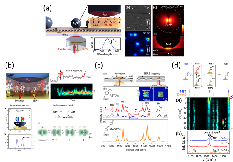
It is well-established that carefully designed plasmonic gaps offer sufficiently large field enhancement for single-molecule level SERS (smSERS). An obvious next step is the application of smSERS for the study of chemical reactions at metallic surfaces. However, typical smSERS signals exhibit spatial heterogeneities and temporal fluctuations, making them difficult to be used in such the studies. We have recently developed an experimental strategy to “tame” the stochastic smSERS signals, and to isolate genuine single-molecule reaction events from the time-trajectories of SERS signals. The SERS studies of chemically reactive molecules also have lead us to discover new insights into plasmon-assisted chemical reactions on metallic surfaces, including the dimerization kinetics of 4-aminobenzenethiols , and the formation of nitro-anion radical intermediates in the first step of reduction of 4-nitrobenzenethiol.
* SERS mechanism beyond classical EM and CHEM mechanism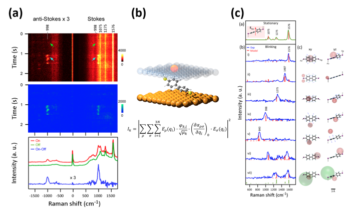
Spectroscopic proof of the existence of atomic-scale SERS hotspots; Shin et al., Nano Lett. (2018).
40 years of investigation on SERS have revealed that the SERS arise from plasmonic field enhancement (so-called the EM-mechanism) and from the chemical (charge-transfer enhancement, perturbation on molecular polarizability, and many others) mechanisms. While the two mechanism overall explain the experiment there still exists many other effects that may be operative. We are exploring such new mechanisms in SERS process. Recently, we have demonstrated that surfaces of metallic structures may possess angstrom-sized hotspots that interact with a single-molecule at angstrom-scale dimensions.
Photons: Nanoscopy
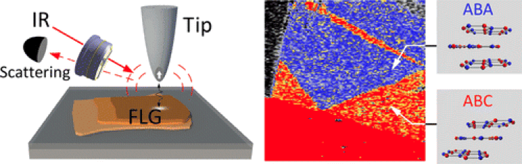
IR nanoscopy technique (left) and image (right) of a trilayer graphene showing ABA and ABC stacking domains; Kim et al., ACS Nano 9,6765 (2015).
In most cases, conventional optical microscopy does not reveal full nano-resolution desired for studying nanostructures and individual molecules. Rather, it only allows us to see images that are significantly blurred. This is called the Abbe's diffraction limit of light. This limit is, however,not a fundamental rule and several techniques exist that evades such limit. We are developing a nanoscopy technique that enables us to image and spectroscopically characterize nano-objects and individual molecules. In particular, we are developing the scattering-type scanning near-field microscopy (s-SNOM) technique in order to achieve nanometric spatial resolution and full spectroscopic information. The biggest advantage of the s-SNOM is that it can be, in principle, applied to far-IR as well as visible radiation. The aim of this project is to push the limit of the s-SNOM to an individual molecule, particularly in IR and FIR regime, which may lead us to the ultimate goal of IR vibrational spectroscopy of single molecules.